Eye Cataract Surgery

What you want to read...
What is Cataract?
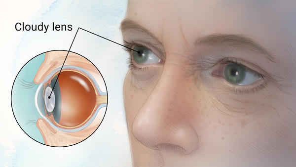
Cataracts refer to clouding of the eye’s natural lens, which is located behind the iris and pupil. The eye’s lens, similar to a camera lens, focuses light onto the retina, located at the back of the eye.
Underlying causes of cataracts in the eye
Cataracts resulting from aging
Cataracts caused by aging are the most common type of cataracts. A significant percentage of people develop this type of cataract, and its prevalence increases with age.
Cataracts resulting from other diseases
This type of cataract is observed in individuals who have specific medical conditions, including diabetes (sugar disease). Sometimes, the occurrence of cataracts is associated with the long-term use of certain medications, such as prednisolone.
Cataracts resulting from injury
Sometimes, cataracts are seen immediately after eye injuries, while in other cases, they may develop years later.
Congenital Cataracts
Some children are born with cataracts, or they develop them in childhood, typically affecting both eyes.
The most common symptoms of cataracts
- Blurriness or vision loss
- Becoming sensitive to light
- Excessive glare from car headlights at night, difficulty with headlight and sunlight glare, and seeing halos around lights.
- Reduced vision in bright sunlight.
- Fading or desaturation of colors.
- Double vision or multiple images (which worsen with increased cataract severity).
- Increased nearsightedness, continuous prescription changes for glasses or contact lenses.
These symptoms can also be caused by other eye conditions, so if you experience any of these symptoms severely, be sure to consult an eye specialist.
Diagnosing cataracts
For a proper diagnosis of cataracts, a comprehensive eye examination by a specialized ophthalmologist is necessary. The examinations performed by an eye specialist for an accurate diagnosis typically include the following:
- Measuring visual acuity
- Examination of the appearance of the eyes
- Eye movement examination
- Examination with a slit lamp device
- Examination of pupil reactions to light
- Tonometry: This examination measures the eye pressure. An increase in pressure can be a sign of glaucoma (high eye pressure).
- Dilation of the pupils, which is done with the use of eye drops, allows the eye doctor to fully examine all parts of the lens and retina
The treatment for cataracts
Cataracts require surgery when they interfere with vision and daily activities like driving, reading, or watching television, and there is no need to wait for them to mature.
In cataract surgery, a small incision of about 3 millimeters is made to remove the cloudy lens. The cataract is broken up within the eye and then removed using suction. This procedure comes in two types:
A-Phacoemulsification
which use ultrasound energy to break up the lens nucleus
B-Femtosecond Laser
It uses “laser” energy to break up the lens nucleus
In Iran, nearly 100% of cataract cases are treated using the “phacoemulsification ultrasound” method, commonly referred to as “phaco.” However, since laser surgery is widely believed to be highly precise, this method is sometimes referred to as the “laser method.”
If both eyes have cataracts, the doctor won’t perform surgery on them simultaneously. Each eye needs to undergo surgery separately.
Regarding anesthesia, with the advancements in surgical technology, there is no longer a need for general anesthesia. The surgery can be performed with local anesthesia, either through anesthesia drops or injections.
The most advanced surgical method in the world is phacoemulsification surgery using local anesthesia. This method doesn’t require general anesthesia or the use of injection anesthesia. The surgery typically takes about ten minutes, and there is no need for eye bandages. Patients are discharged immediately after the surgery and can return to their regular activities and daily life the next day.
Simultaneous treatment of cataracts and presbyopia
In the past, cataract surgery involved removing the cataract and prescribing thick eyeglasses to compensate for the refractive error it caused. Gradually, with the invention of intraocular lenses and the development of precise formulas for calculating the power of these intraocular lenses, people usually no longer needed eyeglasses for distance vision and only required them for near vision or specific tasks like reading or work.
Subsequently, with the invention of new cataract surgery methods such as “phacoemulsification” (often called “phaco”) and methods for correcting astigmatism, refractive errors such as myopia, hyperopia, and astigmatism could be corrected simultaneously during cataract surgery.
Most people over the age of 40 often experience the need for reading glasses or feel that they require them to read and see objects up close. These changes in the eye’s lens are referred to as “presbyopia.”
Preoperative recommendations
- After deciding to undergo cataract surgery and if you intend to use your health insurance for it, it's advisable to visit the relevant insurance office the day before the surgery with a referral letter from your treating physician to obtain the necessary referral documents for the clinic where the surgery will take place.
- If your treating physician deems it necessary, a day or two before the surgery, you should have blood and urine tests conducted at one of the nearby healthcare centers or hospitals. You can then bring the test results to the clinic on the day of the surgery.
- The day before the surgery, take a shower, and it's advisable for gentlemen to shave their faces.
- On the day of the surgery, it's best for women to avoid makeup, especially eye makeup.
- On the day of the surgery, be sure to take all the medications you use for other conditions such as diabetes, high blood pressure, heart problems, etc.
- If your surgery is scheduled in the afternoon shift, have breakfast. If the surgery is in the morning shift, follow your doctor's recommendations for whether or not to have breakfast.
- On the night before the surgery, rest comfortably and try not to stress, as the surgery is straightforward and easy.
- On the day of the surgery, you should arrive at the clinic for admission one hour before the operation.
- After entering the surgery ward, you will change into the appropriate attire for the operating room, and a nurse will administer dilating eye drops into your eyes several times.
- About half an hour later, you will be taken to the operating room along with a nurse.
- After lying down on the operating table in the operating room, you will have numbing eye drops (such as Tetracaine) applied to your eyes several times. These numbing drops will make your eyes completely numb, and you won't feel any pain during the procedure.
Post-cataract surgery instructions and care
- You will be discharged immediately after the surgical procedure. Make sure to get your prescription medications and use your eye drops as instructed. Bring your medications with you for follow-up visits.
- After the surgery, lie on your back and avoid sleeping on the side of the operated eye or on your stomach.
- Stay calm and avoid coughing, sneezing, and straining.
- You can eat immediately after the surgery. There are no specific dietary restrictions, and no special diet is recommended.
- You should usually use corticosteroid eye drops (like betamethasone or prednisolone) every 2 hours and chloramphenicol eye drops every 4 hours.
- If prescribed, take acetazolamide tablets (for lowering eye pressure) every 6 to 8 hours.
- If you feel pain or discomfort, use acetaminophen tablets.
- At night, when you go to bed, there's no need to use eye drops.
- Allow at least 5 to 10 minutes between two eye drops.
- When going to sleep, a shield should be placed over the eye and secured with hypoallergenic tape.
- The shield should remain in place over the eye during sleep for 3 to 4 weeks after the surgery. During the day, use sunglasses to protect your eye, as the operated eye is sensitive to sunlight.
- Increased tearing and some discharge upon waking up from sleep are normal after cataract surgery. This can cause your eyelashes to stick together. You can gently clean this discharge with a clean tissue.
- Postoperative pain can be alleviated by taking acetaminophen. If the pain persists, you should inform your doctor, as it may be due to increased intraocular pressure.
- A sudden decrease in vision should be reported to the doctor.
- You should visit the clinic for an examination the day after the surgery, and multiple follow-up appointments are necessary for a complete recovery.
- Your vision may not be completely clear immediately after the surgery, but it will gradually improve. After about a month, if necessary, you may be prescribed reading glasses for fine tasks.
- Keep in mind that the quality of your vision after the surgery depends on the condition of the retina, the health of the optic nerve, and other eye components, which will gradually improve.
- In cases where the intraocular lens doesn't provide the exact vision correction needed, patients may require reading glasses for activities such as reading. These glasses are typically prescribed about a month after the surgery.
پس از عمل جراحي آب مرواريد نكات زير بايد رعايت شود و تا ويزيت بعدي از انجام كارهاي زير خودداري نماييد.
- استفاده از مواد آرايشي براي چشم و اطراف آن
- شستشو و استحمام چشمها
- ورزش و فعاليتهاي سنگين و بلندكردن اجسام سنگين
- مالش و دست زدن به چشمها
- سرفه كردن و عطسه كردن
- زورزدن در هنگام اجابت مزاج
- تغيير ناگهاني وضعيت سر
- خم شدن سر تا زير كمر و سجده نمودن. به مدت 3 تا 4 هفته در هنگام نمازخواندن به سجده نرويد بلكه مهر را به پيشاني خود بگذاريد.
- خوابيدن به طرف شكم و يا چشم عمل شده
- شستن سر و صورت با شامپو بعد از ويزيت روز اول پس از عمل جراحي و با صلاح پزشك بلامانع است .
- در صورت داشتن بينائي کافي رانندگي و تماشاي تلويزيون و کار با کامپيوتر تا جائي که شما را خسته نکند، بلامانع است.
- بعد از آنكه به شما اجازه استحمام داده شد از ورود آب به داخل چشم به مدت 2 تا3هفته خودداري كنيد.
- در موقع استحمام از مالش چشم ها جداً بپرهيزيد.
